
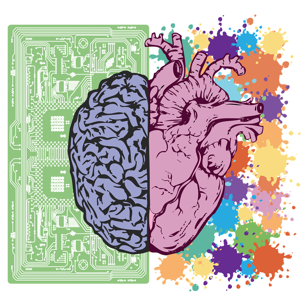
Heart on a chip! Sounds futuristic? Dive deep into the article.
Cardiovascular diseases (CVDs), according to the World Health Organization (WHO), are the leading cause of death globally. A 2019 report revealed that 18 million people from Asia alone have succumbed to CVDs. A primary reason for these numbers is the lack of efficient human-relevant models to study cardiovascular diseases. Meet HoC devices (heart-on-a-chip devices), an innovative platform used to replicate the structure, intricate functions, and dysfunctions (in a diseased condition) of a human heart in vitro. This offers a powerful alternative to conventional models.
Currently used models
Human models
They provide the most direct and realistic data but ethical issues and availability limits their usage.
Animal models
They are easier to study with relatively fewer restrictions. They are useful for understanding disease progression and treatment. However, they are time-consuming. Moreover, physiological differences between animals and humans make them unreliable.
In-vitro models
Using basic 2D cell cultures or animal models may be helpful, but, they have limitations. They fail to capture the complexity of the disease and cannot predict how a human will respond to the particular disease and treatment strategy. A tiny chip-based approach can help address all these shortcomings by giving us a model with high accuracy concerning physiological, anatomical, and bio-mechanical aspects of the heart. This makes it a relevant platform for drug testing, disease modeling, and personalized therapy as it mimics your heart. With high biomimicry, and low ethical issues involved, they are both efficient and economical.
Why heart-on-a-chip matters?
They are miniaturized devices that mimic the physiology, anatomy, and bio-mechanical features of a human heart. This in-vitro model has helped us get a better understanding of diseases like ischemia, myocardial infarction, and other cardiovascular diseases. They offer the following advantages when it comes to CVD models.
- High accuracy
- Low ethical concerns
- Cost-effective
- Enable drug testing, disease modeling, and personalized therapy
Highly advanced HoCs can even provide biomechanical simulations to cardiac cells developing them into fully functional heart issues.
Limitations of current HoC models
- They cannot achieve cardiocyte maturation to become fully functional cardiac cells
- Incomplete replication of electrophysiological properties of the cardiac cells
- Limited ability to mimic mechanical contractions of the heart
- Environmental control remains complex
- The selection of bio-material is tricky
- Integration into drug testing pipelines is still underway
- Scalability and cost-effectiveness need improvement
Building a HoC
Bio-materials
The choice of bio-material is very crucial. It should be bio-compatible, have good optical properties, and thermal conductivity, and affordable. They are useful in creating three-dimensional cell scaffolds that mimic the 3D environment inside our body.
Commonly used materials include-
Polydimethylsiloxane (PDMS) – great breathability, flexibility, and transparency
Polymethyl Methacrylate (PMMA)- suitable for mass production, but lacks oxygen permeability
3D printing models also use Polyethylene Terephthalate (PET), Polycaprolactone (PCL), Acrylonitrile Butadiene Styrene (ABS), and PMMA
Cell adhesion
Once the chip is constructed, cells must adhere to it. Traditional 2D reagents like collagen and fibronectin are used but are not suitable for 3D environments.
To mimic the actual extracellular environment, 3D scaffolds made of PCL, PLA, and hydro-gel composites are preferred.
To enhance electrical conductivity and mimic in-vivo conditions more closely, materials like gold nanoparticles or carbon nanotubes can be added.
Cell sources
Cells used include-
- Primary cardiac cells (from humans and animals)
- Stem cells
- Pluripotent stem cells
- Cardiac muscle cell lines
HL-1 cells naturally contract, making them ideal for modeling arrhythmias or atrial fibrillation. However, using human cardiac cells raises ethical concerns, and stem cells, though versatile, can be expensive and prone to mutations.
Fabrication of an HoC
There are two main categories:
1. Mold-dependent fabrication
Like using a cupcake tray, materials are shaped using a mold, ensuring uniformity and mass production.
2. Mold-independent fabrication
Think of decorating cupcakes freehand like using a piping bag. It is more creative but less scalable. Techniques like 3D printing fall into this category, offering flexibility in design.
Most common fabrication methods include Lithography and organ printing While these techniques are fascinating, diving into their technical details is beyond the scope of this article. But don’t worry, an engaging post on organ printing is coming up soon, so stay tuned if you’re curious about how scientists are “printing” living tissues!
Stimulation of Cardiac Tissue
In vitro, cardiac stem cells are immature and do not exhibit full heart-like behavior. To mimic real heart activity, scientists use two types of stimulations:
- Mechanical stimulation (e.g., a probe applies stress to cells, promoting maturation)
- Electrical stimulation (using electrodes made of carbon or platinum)
These stimuli help the cells develop characteristics similar to those in a living heart.
Detecting Physiological Parameters
Thanks to integrated sensors, HoCs can monitor:
- Electrophysiology- determine the electrical signals controlling the heartbeat. Abnormal electrophysiology can be caused due to cardiac arrhythmia or cardiac arrest. So studying electrophysiology of the heart is very vital and that is now possible thanks to heart on chips. Materials like gold, silver, graphene, and carbon nanotubes are used for their high conductivity and stability.
- Contractility – this tells us how strongly and rhythmically cells contract.
Monitoring contractility
Yes—heart contractions can be seen on a chip!
Using fluorescent microscopy, scientists introduce fluorescent markers into the microfluidic channels. These markers light up during cell contraction, allowing for real-time observation and analysis.
Disease-Specific Heart-on-a-Chip Models
What makes Heart-on-a-Chip technology truly revolutionary is its adaptability.
Researchers can now design HoCs tailored to specific cardiac diseases, allowing us to study conditions like ischemia, myocardial infarction, cardiac fibrosis, cardiac scarring, and myocardial hypertrophy in a controlled, in vitro environment. No animals, No human subjects.
Just science — precise, ethical, and replicable.
As advancements continue, these tiny chips are set to transform how we understand, model, and ultimately treat heart disease.
This is the future of heart research.
Want to see this technology in action?
Dive deeper into the world of Heart-on-a-Chip with our latest video.
Discover how this tiny chip mimics the human heart and why it’s a game-changer in cardiac research. Subscribe to itsbiodiaries and stay tuned for a Heart on Chip part 2.
Stay curious, stay inspired!
Wow, this is the future!
Great read, thanks for sharing.