
Western blotting is a widely used technique in molecular biology to detect a specific protein in a complex sample using probes like antibodies.
Researchers often perform western blotting after SDS-PAGE, which separates proteins based on their molecular weight.
Working Principle
Western blotting relies on the specific interaction between an antigen (protein of interest) and an antibody.
The process involves two main steps:
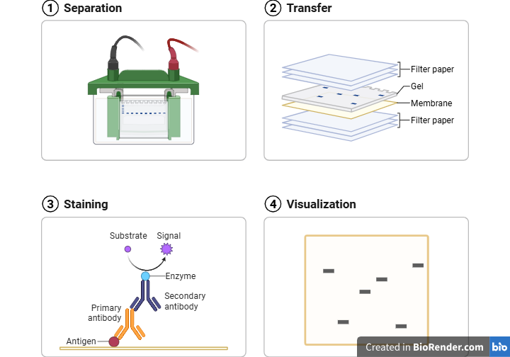
- Separating proteins using SDS-PAGE
- Transferring (blotting) the separated proteins onto a membrane such as:
- Nitrocellulose
- Polyvinylidene fluoride (PVDF)
Once transferred, antibodies bind specifically to the target protein, allowing its detection.
Methods of Detection
You can detect proteins using:
1. Primary Antibody (Direct Method)
A labeled primary antibody binds directly to the target protein.
2. Secondary Antibody (Indirect Method)
An unlabeled primary antibody binds to the protein, followed by a labeled secondary antibody that binds to the primary antibody.
Reporter Systems
The antibody carries a reporter (probe) that helps visualize the protein.
Common reporters include:
- Enzymes that produce a color change
- Enzymes that generate a luminescent or fluorescent signal
These signals appear at the antigen–antibody binding site and require a suitable detection system.
Procedure
1. Sample Preparation and Separation
Prepare the sample and separate proteins using SDS-PAGE.
2. Protein Transfer (Blotting)
Transfer the separated proteins onto a membrane using a transfer buffer (commonly containing 10% methanol).
Assembly of the Transfer Stack (Bottom to Top):
- Foam sponge (pre-wet with transfer buffer)
- Filter paper
- Gel
- Membrane (ensure no air bubbles between gel and membrane)
Place the setup in a transfer tank filled with buffer and apply 100 V for ~1 hour.
3. Immunodetection
a. Washing
Wash the membrane with saline for 5 minutes.
b. Blocking
Immerse the membrane in 10% skimmed milk in Tris buffer for 30 minutes.
This step prevents non-specific binding of antibodies.
c. Washing
Wash the membrane again with Tris buffer.
d. Primary Antibody Incubation
Add the primary antibody and incubate for 3 hours.
Wash the membrane to remove excess antibody.
e. Secondary Antibody Incubation (if used)
Add an HRP-conjugated secondary antibody (1 µg/mL) and incubate for 1 hour.
Wash again to remove unbound antibodies.
f. Detection
Add the substrate and incubate for ~5 minutes before detecting the signal.
Advantages
- Requires small amounts of sample and reagents
- Allows multiple analyses from the same transferred protein sample
Applications
- Detects proteins even in very low concentrations
- Serves as a confirmatory test for HIV
- Helps in protein quantification and analysis of gene expression
Quick Summary
Western blotting combines:
- Protein separation (SDS-PAGE)
- Transfer to a membrane
- Specific antibody-based detection
This makes it a powerful and reliable technique for studying proteins.